
Guide
MRI on iPhone: what you can and cannot see
To view an MRI on a phone, copy the MRI disc or ZIP to your iPhone or iPad and import it into a DICOM viewer like Ossidex. Each MRI sequence appears as its own series; scroll through slices, adjust contrast, and place several series side by side so the reference lines follow as you scroll.
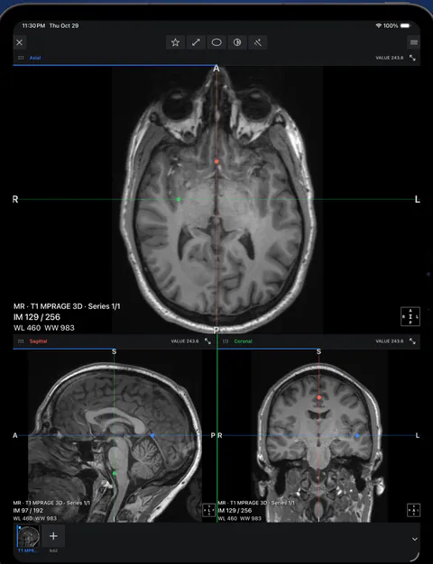 rtAttenPenn · Zenodo 3873446 · CC BY 4.0
Try it on the built-in brain MRI
Since version 2.0 the app includes a brain MRI demo next to the dental CBCT demo: three series of one examination (a sagittal T2, an axial T1 after contrast and an axial FLAIR) with linked reference lines. Pick it right after onboarding to see multi-panel layouts and linked reference lines before you import your own study. In 2.0, 3D for MRI is fast again and starts with a better default window.
View an MRI in Ossidex
- Get the files. Copy the MRI disc folder or ZIP to Files, or keep the download link the imaging centre sent.
- Import. Tap + in Ossidex and choose the folder, ZIP or paste the link.
- Pick a series. Open the study and tap a series thumbnail. Swipe up and down to move through the slices.
- Adjust contrast. Drag on the image to change window/level; MRI has no fixed units like CT, so contrast is adjusted by eye.
- Compare series. Add series to a multi-panel layout. Scroll one and the reference lines show the same position in the others (multi-panel is a Pro feature on your own studies).
What a phone viewer cannot tell you
A viewer displays the series you received; it cannot supply a missing sequence or turn a screenshot into a full examination. Opening an MRI is not the same as interpreting it. Use the radiologist’s report and discuss findings with the clinician who knows the reason for the examination.
In Ossidex 2.0, open the built-in brain MRI demo, select its T1 3D series and explore the linked planes. Then compare that experience with your own imported series: a thick-slice 2D acquisition is not an equivalent 3D volume. Import studies, view slices in 2D and measure on the device. MPR and multi-panel layouts on your own study require Pro; paying for Pro does not add missing slices or spatial information.
How an MRI study is organised
An MRI examination is one study made of many series. Each series is one acquisition — a particular sequence in one plane — with dozens to hundreds of slices. A knee MRI might contain a sagittal proton-density series, a coronal T1 series and an axial fat-suppressed series. Ossidex shows every series as a thumbnail; the series description (for example SAG PD FS) comes from the scanner.
Phone or tablet?
An iPhone is fine for checking a study or showing someone a finding. An iPad is better for longer reading and for comparing several series at once, because every panel stays large enough to see detail. Both run the same app and the same tools.
Planes and slice thickness
Unlike CT, MRI is often acquired directly in the plane the radiologist wants — sagittal for the spine, coronal for the knee — and each plane is a separate series. Slices are frequently 3–5 mm thick with a gap between them, so scrolling looks coarser than CT. Some protocols include a thin 3D acquisition (series names like 3D, MPRAGE, SPACE, CUBE or VISTA); those reconstruct well in MPR. The slice number and total (for example IM 11 / 50) is shown in the corner of each view.
Brain, spine, joints and TMJ
The same viewer works for any body region. Brain studies typically contain axial T1, T2, FLAIR and diffusion series; spine studies sagittal and axial T1/T2; joint studies proton-density series in three planes. Dentists and gnathologists receive MRI of the temporomandibular joint, often with open- and closed-mouth series — open them side by side in a multi-panel layout to compare. Ossidex also opens NIfTI files used in research, including segmentation masks as layers.
Before you rely on it
Scans are opened and processed on your iPhone or iPad. There is no account, and your images are not uploaded to Ossidex servers.
Try it and verify with your study
Start with the built-in sample: MPR, 3D and the arch are unlocked on it. When importing your own study, check series count, orientation and that every file arrived together. Our compatibility matrix distinguishes recorded results from general support claims. Check the App Store terms before choosing.
Details: MRI on iPhone: ZIP import and two series
MRI on iPhone: ZIP import and two series. Import a public MRI ZIP with 416 images, open the study and compare sagittal and derived axial series with reference lines.
Recorded on October 15, 2026: Ossidex 1.11.2, iPhone 17 Pro Simulator, iOS 27.0. A local development build from the released version’s source, with Pro tools enabled. Pauses are edited; there is no voiceover.
Details: Open DICOM in your browser
The browser viewer offers free 2D and MPR, with a linear ruler in millimetres when valid spatial calibration is available. The ruler is unavailable without valid spacing. Angle measurements, ROI, export, 3D and the dental arch are available in the iPhone/iPad app.
Details: Common sequences in plain words
Sequences produce different contrast from the same anatomy. The names below are general orientation, not a guide to interpretation:
| Sequence | General appearance |
|---|---|
| T1-weighted | Fat bright, fluid dark; often used to show anatomy |
| T2-weighted | Fluid bright; commonly used to show fluid and swelling |
| FLAIR | T2-like with the signal from free fluid (e.g. CSF) suppressed; typical in brain imaging |
| PD (proton density) | Common in joint imaging, often with fat suppression |
| DWI / ADC | Diffusion imaging; usually part of brain protocols |
| Post-contrast T1 | Acquired after a contrast agent; labelled “+C”, “POST” or “GD” |
Reading an MRI is a clinical skill. Use the viewer to look at your images or share them with your clinician; the radiologist’s report remains the interpretation.
Details: Which app should I use?
Several iPhone and iPad apps open MRI. The main differences are how the study gets in and whether it stays on the phone (US App Store, September 2026):
| App | Price | Where the MRI is processed | Good for |
|---|---|---|---|
| IMAIOS DICOM Viewer | Free for personal use | On the device | Individual files, also on Android and the web |
| 3DICOM Mobile | Free app; paid platform plans | Loaded from the 3DICOM cloud platform | 3D presentation and sharing |
| Ossidex | 2D, multi-panel, MPR and 3D viewing | On the device, no upload | Discs, ZIPs and download links; linked series |
More apps are compared in best DICOM viewer apps for iPad and iPhone.
Frequently asked questions
Can I open an MRI CD on iPhone?
Not from the disc itself — iPhones have no disc drive. Copy the disc once on a computer or ask for a download link, then import it. Details: how to open a DICOM CD on iPhone or iPad.
Why does my MRI look too dark or too bright?
Change window/level by dragging on the image. MRI signal intensity is relative, so each series needs its own adjustment.
Can I see the MRI in 3D?
Series with thin, contiguous slices can be shown in MPR and 3D (Pro). Many MRI series are acquired as thick slices in one plane, which limits reconstruction.
How can I try the MRI viewing tools?
Ossidex opens studies in 2D, linked MPR, 3D and the dental arch, with measurements and reports. Try the tools on the built-in dental CBCT and brain MRI samples.
How do I open an MRI ZIP file on an iPad?
Save the complete ZIP to Files. In Ossidex, tap +, choose the archive and import it. DICOM sequences appear as separate series. If the archive is incomplete or uses unsupported encryption, ask the clinic for an ordinary ZIP with all images. Archive import guide.
Can I compare MRI series side by side?
Yes. Use a multi-panel layout for series from the same study; reference lines show positions across planes where the DICOM geometry permits. This layout requires Pro on your own studies. It does not automatically register separate examinations. Linked panels and MPR.
Can I view MRI offline and open NIfTI files?
After import, the app reopens your MRI without internet or an account. For research files ending in .nii or .nii.gz, use native NIfTI-1/2 support, including scalar 4D volumes and aligned layers. See NIfTI viewer requirements.
Sources
Related pages
Patients
Got a CD or link with your scan? View it at home, privately.
MPR and 3D volume rendering on iPhone and iPad
Multi-planar reconstruction with linked crosshairs, oblique planes and slab MIP, plus 3D volume rendering with presets and clipping, for CT, CBCT and MRI.
Open a DICOM CD on iPhone or iPad
Open a clinic’s DICOM study on iPhone or iPad from USB, a folder or a link. Learn what to request, how to import it and which viewing tools to use.
NIfTI viewer for iPhone and iPad: open .nii and .nii.gz files
Open NIfTI-1/2 .nii and .nii.gz files on iPhone and iPad: MRI volumes, 4D time points, MPR, label maps and continuous overlays with Ossidex.
View the TMJ on a CBCT scan
Corrected sagittal and coronal planes of the condyle, what CBCT cannot show, and when TMJ MRI is needed.
Ossidex videos: CBCT, MRI, MPR and dental arch
Full CBCT and MRI import walkthroughs on iPhone, plus four shorter iPad demonstrations of MPR, dental arch and 3D.
DICOM export compatibility reference
Compare documented DICOM exports and tested public samples by scanner software: folders, DICOMDIR, codecs and project files. Download the CSV or JSON reference.
Free online DICOM viewer: open CT, MRI and X-ray in your browser, no upload
Open CT, MRI and X-ray DICOM files online for free on Mac, Windows or iPad. Local 2D and MPR viewing, no upload, installation or account.