
Dental CBCT
Dental panoramic arch and cross-sections from any CBCT
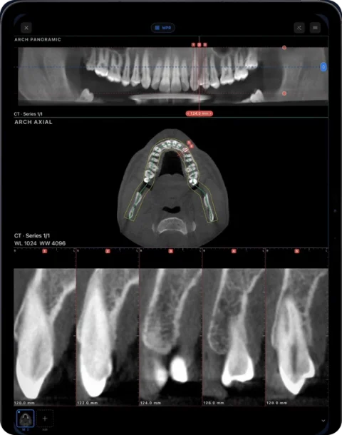
In Ossidex you trace the dental arch on an axial CBCT slice — or let it propose one automatically — and it builds a live panoramic reconstruction with numbered cross-sections perpendicular to the curve. You can measure on the cross-sections, number teeth in FDI or Universal notation and export an arch PDF report.
How it works
- Open the CBCT. Import the study and open it. The axial slice at the level of the teeth is shown first.
- Trace or auto-detect the arch. Tap along the arch, or use Auto: Ossidex proposes a curve based on bone density that you then adjust point by point — move, insert, delete or extend points.
- Read the panorama. A panoramic image reconstructed along the curve updates live while you edit. Change the offset and depth of the curved slab to bring roots or the canal into focus.
- Step through cross-sections. Cross-sections perpendicular to the arch are numbered and shown in a row. Scrub along the panorama to move them; a mini navigator shows where you are.
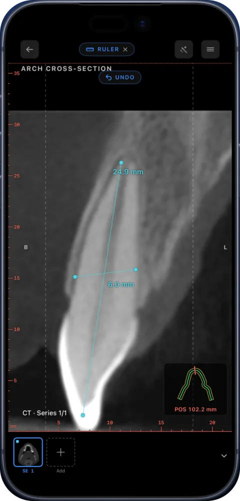
- Measure and export. Measure lengths and angles on a cross-section, read Hounsfield values with the HU probe, and export a PDF with the panorama and all cross-sections.
What dentists use it for
- Measuring bone height and width at a site with the ruler on cross-sections.
- Following a root through thin MPR slabs and measuring root length or canal width with the ruler.
- Locating impacted teeth and their relation to neighbouring roots.
- Explaining findings to patients on the iPad at the chair.
Ossidex is a viewer. It does not design surgical guides or place virtual implants.
Details that save time
- One arch per study: reopening continues where you left off, and measurements move with the curve when you edit it.
- Slice rulers and slice numbers on every cross-section.
- The same colour palette as MPR, so planes are easy to match.
- HU probe with a Misch D1–D4 bone-density reading as a guide (why CBCT values are relative).
Works with every CBCT vendor
The arch is built from the standard DICOM volume, so it works the same for scans exported from Planmeca Romexis, Sidexis, CS 3D Imaging, Ez3D-i, OnDemand3D, NNT and others. See how to export from each vendor.
Panorama from CBCT vs a panoramic X-ray
| Panorama reconstructed from CBCT | Conventional panoramic X-ray (OPG) | |
|---|---|---|
| Source | The 3D CBCT volume | A separate 2D exposure |
| Adjustable after the scan | Yes — curve, offset and depth | No |
| Cross-sections | Yes, perpendicular to the arch | No |
| Measurements | In mm on cross-sections | Limited by magnification and distortion |
More background in panoramic X-ray vs CBCT.
Settings in one card
- Offset moves the curved plane towards the lips or the tongue, to bring roots or the canal into the panorama.
- Depth sets the thickness of the curved slab: thin for detail, thicker for a smoother overview.
- Autofocus centres the curve on the densest structures along the arch.
- Shifting the plane on the panorama is saved and carries existing measurements with it.
Coming soon
- Implant planning Soon
- Endodontic tools Soon
Today you measure bone height and width with the ruler on cross-sections, and follow a root in MPR with a thin slab.
Frequently asked questions
Is the arch available in the free version?
You can try it on the built-in sample CBCT for free. On your own studies it is a Pro feature.
Can it trace the arch automatically?
Yes. Auto proposes an editable curve; you confirm or adjust it.
Does it show the mandibular canal?
You can see the canal on the panorama and cross-sections and measure distances to it. There is no automatic canal tracing.
Does it work on iPhone?
Yes, though the iPad’s larger screen is more comfortable for the panorama plus cross-sections.
Can I export the panorama?
Yes — the arch PDF report contains the panorama and every cross-section.
Related pages
Dental CBCT viewer
Panoramic arch, cross-sections, MPR and 3D for cone beam CT from any scanner.
Reviewing a CBCT before an implant
How to review a CBCT at a planned implant site: arch, cross-sections, ridge height and width, canal and sinus proximity. Ossidex is a viewer only.
How to read a CBCT scan
Navigate a dental CBCT: axial, coronal and sagittal planes, panoramic reconstruction, cross-sections, window/level and common artifacts explained.
DICOM measurements and PDF reports on iPhone and iPad
Measure length, angles and Hounsfield units on CT, CBCT and MRI in Ossidex, annotate the view, and export an image or a one-page PDF report, anonymized.
How to show a patient their scan on an iPad during a consultation
A practical workflow for explaining a CT, MRI or dental CBCT to a patient on an iPad: get the study onto the device, pick the views, measure, share safely.