
Dental X-ray
Panoramic X-ray viewer for iPhone and iPad
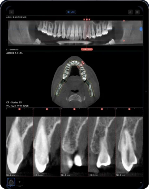
Ossidex (formerly MedScan), a DICOM viewer app for iPhone and iPad, opens dental panoramic radiographs (OPG), periapical and cephalometric X-rays stored as DICOM, with zoom, window/level and measurements — no laptop needed. If you have a CBCT instead, its arch view reconstructs a panoramic image along the dental arch with cross-sections.
2D dental X-rays in DICOM
Panoramic units and intraoral sensors store images in DICOM when the practice software exports them that way, and imaging centres usually hand them over as DICOM files on a disc or through a link. Ossidex recognises these single images alongside CT and CBCT and shows them full-width:
- Panoramic radiographs (OPG / OPT): the whole dentition, both jaws, the condyles and the sinuses in one image.
- Periapical and bitewing images: intraoral films from digital sensors or phosphor plates, exported as DICOM.
- Cephalometric radiographs: lateral and frontal cephs used in orthodontics, which you can measure with the ruler and angle tools.
Zoom and pan with gestures, change brightness and contrast with window/level, and add length, angle, arrow and text annotations. Export the annotated image or a PDF report.
A panorama from a CBCT
A CBCT is a volume, not a single image, but you often want the familiar panoramic overview. In the arch view you trace a curve along the dental arch on an axial slice (or adjust the curve Ossidex proposes), and the app reconstructs a panoramic image along that curve. Unlike a conventional OPG, this panorama has no magnification differences or ghost images from the opposite side, and you can change its depth and thickness and scroll cross-sections at any point along the arch. More on the arch view and panoramic X-ray vs CBCT.
Getting the images onto your phone
Ask for the images in DICOM format. JPEG or PNG exports from practice software lose the pixel spacing Ossidex needs for measurements in millimetres. Then import the files from the Files app, AirDrop, email, a messenger or a cloud link (link import). If the images arrive on a disc, copy the whole disc folder first — see opening a DICOM CD.
Measurements on a conventional panoramic radiograph are affected by the machine’s magnification and patient positioning, and the DICOM calibration may not reflect true anatomical size. Use CBCT for dimensions you rely on.
Dental image types Ossidex opens
| Image | DICOM modality | What you can do |
|---|---|---|
| Panoramic (OPG) | PX | Zoom across the full width, window/level, length and angle measurements, tooth markers |
| Periapical and bitewing | IO | Zoom, contrast, measurements, annotations |
| Cephalometric | DX / CR | Lengths and angles for orientation, annotations |
| CBCT | CT | MPR, 3D and the panoramic arch with cross-sections (Pro) |
Photos attached to a study (JPEG, HEIF) and PDF documents such as a radiologist’s report are kept with the study they came with.
Measurements on 2D X-rays
On a panoramic image, magnification differs horizontally and vertically and changes across the image, so millimetre values from an OPG are approximate even when the DICOM file carries a pixel spacing. Use them for orientation and comparison rather than exact sizes. On periapical images taken with a paralleling technique, distortion is smaller. When a precise distance matters, a CBCT cross-section is the better source — its geometry is the same in every direction.
Opening a panoramic X-ray step by step
- Ask for DICOM. Request the image as DICOM (.dcm) rather than JPEG — practice software can usually export both.
- Save it to Files. From email or a messenger, use Share → Save to Files, or copy the export folder from a USB stick.
- Import in Ossidex. Tap +, choose Files and select the file or folder; the image appears under the patient.
- Adjust and annotate. Pinch to zoom, drag with two fingers for contrast, add tooth markers or arrows, and export an image or PDF if you need to share it.
Frequently asked questions
Can I open a JPEG of a panoramic X-ray?
Ossidex is built for DICOM. Photos and JPEG files attached to a DICOM study are shown with that study, but for measurements in millimetres ask for a DICOM export.
Is viewing 2D X-rays free?
Yes. 2D viewing, window/level and measurements are free. The arch reconstruction from CBCT is part of Pro.
Can I compare an OPG and a CBCT of the same patient?
Yes. Both open in the same patient in your library, and Pro multi-panel layouts show series side by side.
Related pages
Dental CBCT viewer
Panoramic arch, cross-sections, MPR and 3D for cone beam CT from any scanner.
Panoramic X-ray vs CBCT
How a dental panoramic X-ray (OPG) differs from a CBCT: 2D projection vs 3D volume, distortion and magnification, and what each is typically used for.
DICOM measurements and PDF reports on iPhone and iPad
Measure length, angles and Hounsfield units on CT, CBCT and MRI in Ossidex, annotate the view, and export an image or a one-page PDF report, anonymized.
Dentists
CBCT from any scanner at the chair: arch, cross-sections, MPR, 3D and TMJ MRI.