
Guide
How to view the TMJ on a CBCT scan — and when you need MRI
A CBCT shows the bony parts of the temporomandibular joint (TMJ) in three dimensions: the condyle, the glenoid fossa, the articular eminence and the joint space between them. It does not show the articular disc or joint fluid — for those you need an MRI, which Ossidex also opens. First check that the field of view includes both condyles, then turn the MPR planes to follow each condyle’s long axis and scroll through it slice by slice.
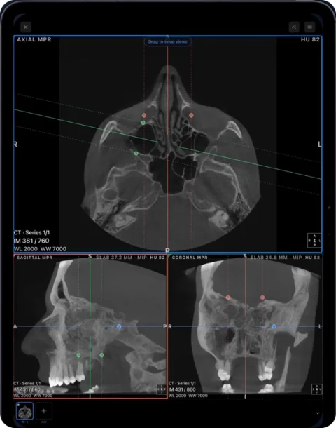
Does your CBCT include the joints?
The AAOMR/AAOP position statement on TMJ imaging asks that the field of view cover at least the condyle, glenoid fossa and articular eminence, and that both joints be imaged, because many TMJ conditions are bilateral. A small field of view taken for a few teeth may not reach the joints at all: scroll the axial slices up from the teeth and check that both condyles and their fossae are complete. The statement also recommends scanning the joints with the teeth together; if the patient bit on a positioning block, keep that in mind before judging condylar position.
Step by step: the TMJ in Ossidex
- Open MPR. Import the study, open the thin-slice CBCT series and switch to MPR: axial, sagittal and coronal planes with linked crosshairs (Pro on your own studies; try the gestures on the built-in sample CBCT).
- Find the condyle on axial. Scroll to the level where the condylar head is widest from side to side and put the crosshair in its centre.
- Align the planes with the condyle. The condyle’s long axis runs obliquely, so standard planes cut it at an angle. Rotate the reference lines on the axial view so that one runs along the long axis and the other crosses it at a right angle: the plane along the axis is the corrected coronal, the plane across it the corrected sagittal.
- Keep the slab thin. Leave slab thickness at its minimum to see the cortical outline; a thick MIP slab projects neighbouring bone into one image and can hide small surface changes.
- Scroll through the condyle. Step through the corrected sagittal from the lateral to the medial pole, then the corrected coronal from front to back. A change may show on only a few slices.
- Measure both sides the same way. On the central corrected sagittal slice, use the ruler for the anterior, superior and posterior joint space, and the angle tool for slopes such as the articular eminence. Repeat every step on the other condyle.
- Check the shape in 3D. Use the bone preset and the clipping plane to see condyle shape and symmetry. Thin cortical bone can fade out of a 3D view and leave holes that are not real, so judge surfaces on the slices.
What Ossidex does not do: there is no automatic TMJ protocol. You orient each condyle by hand, measure joint spaces manually, and the two sides are not laid out side by side for you.
Terms you will see in a TMJ report
Reports usually follow the image analysis criteria developed for the Research Diagnostic Criteria for TMD (Ahmad et al., 2009):
| Term | What it describes |
|---|---|
| Flattening | A flat area on the normally rounded condyle, fossa or eminence |
| Subcortical sclerosis | A denser, brighter band of bone under the joint surface |
| Erosion | A break in the cortical outline |
| Osteophyte | A bony outgrowth at the margin, often on the front of the condyle |
| Subcortical cyst | A rounded dark area under the surface — not a true cyst |
| Condylar position | Condyle concentric, anterior or posterior in the fossa, judged from the joint spaces |
In those criteria, erosion, subcortical cysts, osteophytes and generalised sclerosis indicate osteoarthritis, while flattening or localised sclerosis alone is graded indeterminate and often read as remodelling. A narrow joint space or a posterior condyle is sometimes taken as an indirect sign of disc displacement, but CBCT does not show the disc, and non-concentric condyles are common in people without symptoms.
Interpretation belongs to a trained clinician. This table explains words in a report; it is not a diagnostic checklist. The radiologist’s report remains the interpretation, and Ossidex displays and measures images without interpreting them.
TMJ MRI: the disc and joint fluid
MRI is the only imaging method that reliably shows where the articular disc is. For suspected disc displacement the AAOMR/AAOP statement recommends proton-density or T1-weighted images with the mouth closed and open, in corrected sagittal and coronal planes, plus T2-weighted images when joint effusion is suspected; neither CBCT nor medical CT shows effusion. The open-mouth images show whether a displaced disc returns to its place and how far the condyle moves.
In Ossidex each acquisition is its own series. Place the closed-mouth, open-mouth and T2 series side by side in a multi-panel layout (Pro) and scroll each to the same joint. There is no TMJ-specific MRI tool: Ossidex does not pair open- and closed-mouth slices or mark the disc. Many TMJ series are 2D with a few slices per joint, so read them in their own plane; MPR needs a volumetric series.
Which view answers which question
| Question | Best view |
|---|---|
| Condyle outline, erosions, osteophytes | CBCT corrected sagittal |
| Medial and lateral poles, condyle width | CBCT corrected coronal |
| Orientation of each condyle, left–right comparison | CBCT axial |
| Overall shape, asymmetry | CBCT 3D, confirmed on slices |
| Joint space, condylar position | CBCT corrected sagittal, teeth together |
| Disc position, displacement with or without reduction | MRI, closed and open mouth |
| Joint fluid (effusion) | MRI, T2-weighted |
A panoramic X-ray shows both condyles as part of the overview, but it distorts their shape and misses many bony changes, especially on the front of the condyle. More in panoramic X-ray vs CBCT and the panoramic X-ray viewer.
Imaging facts checked against the sources below on September 29, 2026.
Frequently asked questions
Can a panoramic X-ray show the TMJ?
It shows both condyles and can be a first look, but it distorts their shape and has low sensitivity for bony changes. CBCT shows the bone in three dimensions; MRI shows the disc.
Does my CBCT include the TMJ?
Only if the field of view reaches the condyles. Scroll the axial slices up from the teeth: both condyles should be visible in their fossae.
Can I see the disc on CBCT?
No. CBCT shows neither the disc nor joint fluid. Disc position needs MRI.
Can Ossidex compare open- and closed-mouth MRI automatically?
No. Open both series side by side in a multi-panel layout and choose matching slices yourself.
Is the free version enough?
The download is free: you can import any study, scroll it in 2D, adjust window/level and measure, and a built-in sample CBCT unlocks every tool so you can try them. MPR, 3D, the panoramic arch, multi-panel layouts and unlimited export are part of Pro, an optional auto-renewing subscription priced in the App Store for your country. Corrected TMJ planes and multi-panel MRI need Pro on your own studies.
Sources
- Mallya SM et al. Recommendations for Imaging of the Temporomandibular Joint. AAOMR/AAOP position statement. J Oral Facial Pain Headache 2023 (checked September 29, 2026)
- Ahmad M et al. RDC/TMD: development of image analysis criteria and examiner reliability for image analysis. Oral Surg Oral Med Oral Pathol Oral Radiol Endod 2009
- Ottersen MK et al. CBCT characteristics and interpretation challenges of TMJ osteoarthritis in a hand osteoarthritis cohort. Dentomaxillofac Radiol 2019
- Bianchi J et al. Radiographic interpretation using high-resolution CBCT to diagnose degenerative TMJ disease. PLoS One 2021
- Paknahad M et al. CBCT assessment of mandibular condylar position in patients with TMJ dysfunction and in healthy subjects. Int J Dent 2015
- Guo D et al. Orientation normalization algorithm for the mandibular condyle in small field-of-view CBCT. Dentomaxillofac Radiol 2025
- Verhelst PJ et al. Validation of a 3D CBCT-based protocol for the follow-up of mandibular condyle remodeling. Dentomaxillofac Radiol 2020
- Venkatesh E, Elluru SV. Cone beam computed tomography: basics and applications in dentistry. J Istanb Univ Fac Dent 2017
- Muneeb M et al. CBCT-based analysis of the anatomic characteristics of the articular eminence. J Craniofac Surg 2025
Related pages
Dental CBCT viewer
Panoramic arch, cross-sections, MPR and 3D for cone beam CT from any scanner.
How to read a CBCT scan
Navigate a dental CBCT: axial, coronal and sagittal planes, panoramic reconstruction, cross-sections, window/level and common artifacts explained.
MPR and 3D volume rendering on iPhone and iPad
Multi-planar reconstruction with linked crosshairs, oblique planes and slab MIP, plus 3D volume rendering with presets and clipping, for CT, CBCT and MRI.
View MRI scans on your phone
View MRI scans on iPhone or iPad: import the disc or link, understand series and sequences like T1, T2 and FLAIR, link windows and adjust contrast in Ossidex.
Dentists
CBCT from any scanner at the chair: arch, cross-sections, MPR, 3D and TMJ MRI.