
Guide
Panoramic X-ray vs CBCT: what each shows
A panoramic X-ray (OPG) is a single 2D image of both jaws, with overlapping structures and uneven magnification. A CBCT is a 3D volume you can slice in any plane, including a reconstructed panorama and cross-sections. Panoramic images give a quick overview; CBCT shows depth and bucco-lingual dimensions when a clinician needs them.
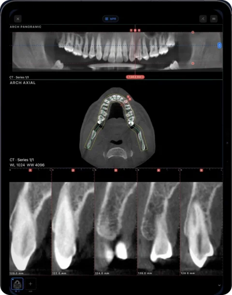
Side by side
| Panoramic X-ray (OPG) | CBCT | |
|---|---|---|
| Image type | One 2D projection | 3D volume of voxels |
| Planes | Fixed curved layer | Any plane: axial, coronal, sagittal, oblique, panorama, cross-sections |
| Geometry | Magnification and distortion vary across the image | Measurable geometry from voxel spacing |
| Overlap | Structures outside the focal trough blur or superimpose | Structures separated in 3D |
| Radiation | Lower than CBCT for comparable exams | Higher; depends strongly on field of view and settings |
| DICOM | Single image (DX/PX) | Multi-frame file or series of slices |
What a panoramic X-ray is good at
An OPG shows all teeth, both jaws, the sinuses’ floor and the TMJ region in one image. It is fast, widely available and a common screening overview. Its limits come from the projection: it cannot show bucco-lingual width, and positioning affects magnification, so lengths on an OPG are approximate.
What CBCT adds
Because CBCT is volumetric, a clinician can scroll through the anatomy, measure width and height, and see relationships in three dimensions. Dental viewers reconstruct a panorama from the volume and add cross-sections, which is why CBCT is often requested for implant assessment, impacted teeth, endodontic questions or pathology. The decision to take a CBCT is clinical, balancing the expected benefit against radiation dose.
Reading a CBCT panorama differently
A panorama reconstructed from CBCT looks familiar but behaves differently. Its thickness can be increased to resemble a conventional OPG or reduced to a thin curved slice; moving the curve buccally or lingually changes what appears. Because it is computed from the volume, the magnification is uniform along the curve, and cross-sections at any point give the bucco-lingual view an OPG cannot. On the other hand, metal restorations produce streak artifacts in CBCT that a panoramic X-ray does not show in the same way.
Other dental X-ray types
| Image | What it is | In DICOM |
|---|---|---|
| Periapical | Intraoral film of a few teeth and their roots | Single 2D image (IO) |
| Bitewing | Intraoral image of crowns of upper and lower teeth | Single 2D image (IO) |
| Cephalometric | Lateral skull projection used in orthodontics | Single 2D image (DX) |
| Panoramic (OPG) | Curved-layer projection of both jaws | Single 2D image (PX) |
| CBCT | 3D volume | Multi-frame or slice series (CT) |
All of these can arrive as DICOM from dental software and open in the same viewer.
Opening both in Ossidex
Ossidex opens panoramic, periapical and cephalometric X-rays as 2D DICOM images and CBCT as a volume. On a CBCT you can build a panorama along the arch with numbered cross-sections (Pro). See the panoramic X-ray viewer and CBCT viewer pages.
How the images are made
A panoramic machine rotates a narrow X-ray beam and detector around the head so that one curved layer — the “focal trough” — is sharp and everything in front of or behind it is blurred. The result is a single flat image of both jaws. A CBCT unit rotates a cone-shaped beam once around the head and reconstructs a 3D block of small cubes (voxels). Any flat view, including a panorama, is then calculated from that block.
This explains the main practical difference: on a panoramic X-ray, anything outside the focal trough is distorted or blurred, and horizontal and vertical magnification differ. On a CBCT, the geometry is consistent in all directions, so a structure can be followed slice by slice and measured on a cross-section.
Questions each image answers
| Question | Panoramic X-ray | CBCT |
|---|---|---|
| Overview of all teeth and both jaws | Yes — its main purpose | Yes, via a reconstructed panorama |
| Bucco-lingual (cheek-to-tongue) width of bone | No — it is a projection | Yes, on cross-sections |
| Position of an impacted tooth relative to roots and canal | Approximate | Three-dimensional |
| Sinus floor and its relation to roots | Approximate | Visible in coronal and sagittal planes |
| Fine detail of enamel and small caries | Limited | Often limited by metal artefacts and voxel size |
The choice of examination is a clinical decision made by the dentist or radiologist; this table only describes what the image types can show.
Looking at both on an iPad
Many patients arrive with an older panoramic X-ray and a new CBCT. In Ossidex you can import both into the same patient, open the OPG as a 2D image and the CBCT as a volume, and put them side by side in a multi-panel layout (Pro). Building the arch on the CBCT gives a panorama with the same overall layout as the OPG, so you can relate the two; the cross-sections then add the third dimension.
Frequently asked questions
Can a CBCT replace a panoramic X-ray?
A CBCT can produce a panoramic-like view, but it involves more radiation and is not used routinely as a replacement. The choice is the clinician’s.
Why are measurements on an OPG unreliable?
Magnification differs across the image and depends on patient positioning, so distances are approximate.
Can I view my OPG on a phone?
Yes, if you have it as a DICOM or image file. Import it in Ossidex like any other study.
Related pages
Panoramic X-ray viewer
OPG, periapical and cephalometric DICOM X-rays, plus panoramas reconstructed from CBCT.
Dental CBCT viewer
Panoramic arch, cross-sections, MPR and 3D for cone beam CT from any scanner.
How to read a CBCT scan
Navigate a dental CBCT: axial, coronal and sagittal planes, panoramic reconstruction, cross-sections, window/level and common artifacts explained.
Dentists
CBCT from any scanner at the chair: arch, cross-sections, MPR, 3D and TMJ MRI.
View the TMJ on a CBCT scan
Corrected sagittal and coronal planes of the condyle, what CBCT cannot show, and when TMJ MRI is needed.