
Guide
CBCT planes explained: axial, coronal, sagittal and the panoramic curve
Reading a CBCT starts with orientation: the axial plane (from above), coronal (from the front) and sagittal (from the side). Dental viewers add a panoramic reconstruction along the arch and cross-sections perpendicular to it. Adjust contrast, scroll systematically and watch for artifacts. Clinical interpretation belongs to a qualified clinician.
What are the CBCT planes?
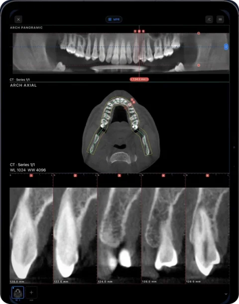
The three CBCT planes are axial, coronal and sagittal views through the same volume. Axial slices show a horizontal cross-section, like looking through the head from above or below. Coronal slices show the front-facing plane, while sagittal slices show the side-facing plane. A panoramic reconstruction follows a curve traced along the dental arch on an axial slice, with cross-sections perpendicular to that curve. Linked MPR views help you navigate between these planes; interpreting findings requires a qualified clinician.
What CBCT is
Cone-beam computed tomography acquires a volume in a single rotation with a cone-shaped X-ray beam. The result is a stack of thin, usually isotropic voxels — the same resolution in every direction — which makes it well suited to reformatting in any plane. It is widely used in dentistry, oral and maxillofacial surgery and ENT. The data is exported as DICOM, often as a single multi-frame file or hundreds of single-slice files.
The three orthogonal planes
| Plane | View from | Typically useful for |
|---|---|---|
| Axial | Above (feet-to-head convention varies by viewer) | Arch shape, bucco-lingual extent, tracing the arch |
| Coronal | The front | Sinuses, left–right symmetry, vertical relationships |
| Sagittal | The side | Anterior–posterior relationships, airway, TMJ region |
In MPR the three planes are linked: moving the crosshair in one updates the others. Orientation labels (A/P, R/L, S/I) at the edges of each view tell you which way you are looking; check them before judging left and right.
Panorama and cross-sections
Because teeth sit on a curve, dental viewers reconstruct a panoramic view along a curve traced over the arch on an axial slice. Unlike a conventional panoramic X-ray it is built from the 3D volume, so its thickness and depth can be changed. Cross-sections are thin slices perpendicular to the curve at regular intervals; they show each site bucco-lingually. In Ossidex you trace or edit the arch, the panorama updates live, and numbered cross-sections run along it with tooth numbering.
A systematic way to scroll
Structured review helps avoid missing areas. A common approach is:
- Confirm patient, date and series, and check the field of view covers the region of interest.
- Scroll the whole volume in axial first, top to bottom.
- Review coronal and sagittal, then the panorama.
- Examine the specific sites of interest with cross-sections and measurements.
- Look at structures at the edges of the field of view as well as the centre.
This is an outline of navigation, not a diagnostic protocol.
Window, level and gray values
Changing window/level alters contrast so that bone, teeth or soft tissue are easier to see. CBCT gray values are not calibrated Hounsfield units like medical CT; they vary with the scanner, field of view and position in the volume. Treat them as relative. See Misch bone density and Hounsfield units.
Common artifacts
| Artifact | Looks like | Cause |
|---|---|---|
| Streaks / beam hardening | Dark and bright streaks radiating from metal | Crowns, implants, amalgam, orthodontic appliances |
| Motion | Double contours, blur | Patient movement during the rotation |
| Noise | Grainy image | Low dose settings or small voxels |
| Cone-beam / edge effects | Uneven brightness near the edges | Geometry of the cone-shaped beam |
Artifacts can mimic or hide findings, which is one reason interpretation requires training.
Doing this in Ossidex
Ossidex opens CBCT from Planmeca, Carestream, Vatech, Dentsply Sirona, NewTom and other units that export DICOM, on iPhone and iPad. Use 2D scrolling, MPR, 3D and the panoramic arch to review a compatible CBCT. Try every tool on the built-in samples.
Frequently asked questions
Is a CBCT panorama the same as a panoramic X-ray?
No. A CBCT panorama is reconstructed from the 3D volume and can be adjusted; a panoramic X-ray is a single 2D projection. See panoramic X-ray vs CBCT.
Why do left and right look swapped?
Medical images are usually displayed as if you face the patient, so the patient’s right is on the left of the screen. Check the R/L labels.
Can I read a CBCT on an iPhone?
You can navigate it on an iPhone; an iPad gives more room for panorama plus cross-sections.
Who should interpret a CBCT?
A qualified clinician. Viewer software, including Ossidex, displays images; it does not interpret them.
Sources
Related pages
Dental CBCT viewer
Panoramic arch, cross-sections, MPR and 3D for cone beam CT from any scanner.
Dental panoramic arch and cross-sections from any CBCT
Trace the dental arch on a CBCT and get a live panorama with numbered cross-sections, tooth numbering, HU probe and a PDF report — on iPad and iPhone.
Panoramic X-ray vs CBCT
How a dental panoramic X-ray (OPG) differs from a CBCT: 2D projection vs 3D volume, distortion and magnification, and what each is typically used for.
Dentists
CBCT from any scanner at the chair: arch, cross-sections, MPR, 3D and TMJ MRI.
View the TMJ on a CBCT scan
Corrected sagittal and coronal planes of the condyle, what CBCT cannot show, and when TMJ MRI is needed.