
Guide
Reviewing a CBCT before an implant: what to look at
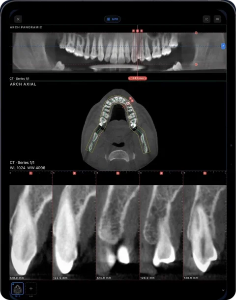
Reviewing a CBCT before an implant usually means tracing the arch, opening cross-sections at the planned site, measuring available bone height and width, and visually checking the distance to the mandibular canal, sinus floor and neighbouring roots. Ossidex supports this viewing on iPad and iPhone; it is not implant-planning or surgical-guide software.
What this page is — and is not
This is a description of how clinicians commonly view a CBCT in a viewer. It is not clinical guidance. Ossidex does not place virtual implants, design surgical guides or recommend treatment.
A typical viewing workflow
- Open the right series. Import the CBCT and check patient, date and that the field of view covers the site.
- Trace the arch. On an axial slice through the alveolar ridge, trace or accept the automatic arch; the panorama builds along it.
- Go to the site. Move to the tooth position on the panorama; tooth numbering (FDI or Universal) helps orientation.
- Read the cross-sections. Scroll the perpendicular cross-sections through the site to see the bucco-lingual profile of the ridge.
- Measure. Use the ruler on the cross-section for height and width of the ridge. Measurements are geometric distances from the DICOM voxel spacing.
- Check neighbouring structures. Look at the canal, mental foramen region, sinus floor and adjacent roots in cross-sections, panorama and MPR.
Structures people commonly review
| Region | Structures | Where to look |
|---|---|---|
| Posterior mandible | Mandibular canal, mental foramen region | Cross-sections and panorama |
| Posterior maxilla | Sinus floor, septa | Cross-sections, coronal |
| Anterior maxilla | Incisive canal, labial plate | Cross-sections, sagittal |
| Any site | Adjacent roots, ridge shape | Panorama, axial |
Visibility of the canal varies between scans. Where a structure is not clearly visible, that uncertainty is itself important.
Measurement caveats
- Measure on a true cross-section perpendicular to the arch, not an oblique cut.
- Voxel size, noise and metal artifacts limit precision.
- Gray values are relative on CBCT; see Misch classes and HU.
- Measurements carry over when you adjust the arch in Ossidex, but re-check them after edits.
Before you start: scan quality
How much a CBCT can show at an implant site depends on the acquisition: field of view, voxel size, patient movement and metal nearby. A small field of view with small voxels shows fine detail but may not include the whole arch or the sinus. Scatter from crowns and existing implants can obscure the ridge next to them. Checking these factors first tells you how much confidence the images support.
It also helps to confirm the orientation: a tilted head produces oblique slices, and the arch curve should follow the ridge at the level you are interested in. In Ossidex the automatic arch can be edited point by point, and the plane offset and depth can be adjusted from one settings card.
Showing the patient
An iPad at the chair lets you show the panorama and cross-sections during a consultation. Ossidex can export the arch view with cross-sections as a PDF report (Pro).
Measuring height and width on a cross-section
- Trace the arch. On the axial slice at the level of the ridge, trace the arch or accept the automatic curve and adjust its points.
- Move to the site. Scrub along the panorama until the numbered cross-section runs through the site you are reviewing.
- Measure height. Use the ruler from the crest of the ridge to the structure you want to keep clear of — for example the canal or the sinus floor.
- Measure width. Draw a second line across the ridge at the levels you care about; repeat on neighbouring cross-sections.
- Check other planes. Confirm the site in MPR and the 3D view, and export an arch PDF with the panorama and cross-sections if you need to share it.
What is coming
- Implant planning Soon
Today Ossidex gives you the viewing and measurement steps above. Virtual implant placement, implant libraries and surgical guides are not part of the current app.
Frequently asked questions
Can Ossidex plan implants?
No. It is a viewer: you can trace the arch, read cross-sections and measure, but it does not place implants or make surgical guides.
Can I show the mandibular canal?
You can review the canal visually in cross-sections, panorama and MPR. Ossidex does not trace or segment the nerve canal.
Is the arch available for free?
The panoramic arch is a Pro feature on your own scans. You can try it free on the built-in sample CBCT.
Related pages
Dental panoramic arch and cross-sections from any CBCT
Trace the dental arch on a CBCT and get a live panorama with numbered cross-sections, tooth numbering, HU probe and a PDF report — on iPad and iPhone.
How to read a CBCT scan
Navigate a dental CBCT: axial, coronal and sagittal planes, panoramic reconstruction, cross-sections, window/level and common artifacts explained.
Misch bone density D1–D4
The Misch D1–D4 bone density classes, the Hounsfield ranges usually quoted for them, and why CBCT gray values are only relative, not true HU.
Dentists
CBCT from any scanner at the chair: arch, cross-sections, MPR, 3D and TMJ MRI.