
Guide
How to view CT scans on iPhone and iPad
To view a CT scan on iPhone or iPad, import the disc folder, ZIP or share link into Ossidex. CT values are shown in Hounsfield units, so window presets for bone, soft tissue or lung give the right contrast instantly; scroll the axial slices, then switch to MPR or 3D to see the anatomy in other planes.
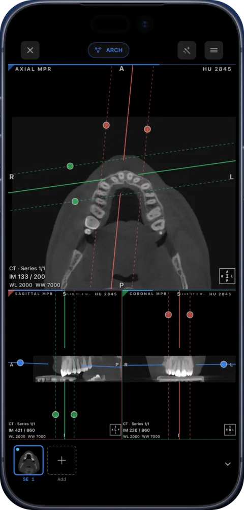 rtAttenPenn · Zenodo 3873446 · CC BY 4.0
Open DICOM in your browser
The browser viewer offers free 2D and MPR, with a linear ruler in millimetres when valid spatial calibration is available. The ruler is unavailable without valid spacing. Angle measurements, ROI, export, 3D and the dental arch are available in the iPhone/iPad app.
Watch the app workflow
An excerpt from a product recording. See all videos and descriptions. Controls may differ from the current release.
Hounsfield units in one paragraph
CT measures how strongly each voxel attenuates X-rays and stores it on the Hounsfield scale, where water is 0 HU and air about −1000 HU. Fat is negative, soft tissue slightly positive, and dense bone reaches hundreds to over a thousand HU. Because the scale is calibrated, Ossidex applies the rescale values in the DICOM header and shows true HU under your finger with the HU probe or an ROI.
Window presets
No screen can show 3000+ HU levels at once, so a viewer maps a window (range) around a level (centre) to grey. Typical starting points:
| Preset | Typical level / width (HU) | Shows |
|---|---|---|
| Soft tissue | 40 / 400 | Organs, muscle, fluid |
| Lung | −600 / 1500 | Lung parenchyma and airways |
| Bone | 400 / 1800 | Cortical and trabecular bone |
| Brain | 40 / 80 | Grey and white matter |
Exact values vary between institutions. Drag on the image to fine-tune.
View a CT in Ossidex
- Import. Tap + and choose the disc folder, ZIP or DICOMDIR in Files, or paste a cloud link.
- Open the series. Choose the study and the series — thin-slice series are best for reconstruction.
- Pick a window. Choose a preset such as bone or soft tissue, then drag to adjust.
- Measure. Use the ruler for distances, the angle tool, or an ROI for mean and range of HU.
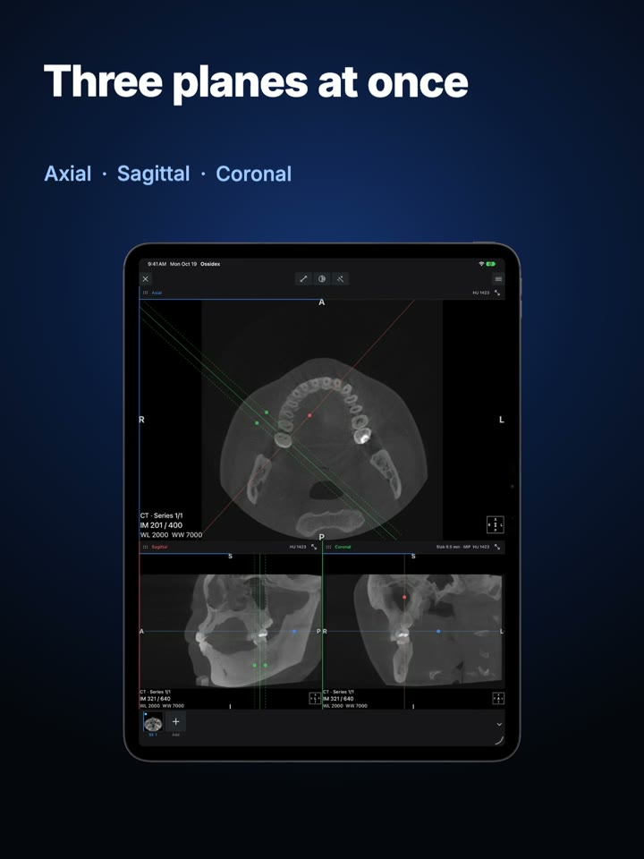
- Reconstruct. Switch to MPR for sagittal and coronal planes, or 3D for a volume rendering (Pro on your own studies).
Which series to open
A CT study often has several series: a scout (localiser), thick-slice reconstructions for quick reading, and thin-slice series for MPR and 3D. Series descriptions such as “1.0 mm bone” or “5 mm soft tissue” come from the scanner. For reconstruction choose the thinnest series.
Reading a CT systematically
Radiologists use structured search patterns so that nothing is skipped. As a viewer user you can borrow the idea: scroll every slice from top to bottom once in a soft-tissue window, then again in a bone window, and for chest studies once more in a lung window. Changing the window changes what is visible — a finding can be invisible in one window and obvious in another. Compare left and right sides at the same level, and use MPR to confirm in a second plane anything you notice in the axial slices.
For contrast-enhanced studies, series labelled “arterial”, “portal venous” or “delayed” were acquired at different times after injection; they are separate series in the study list.
Sharing what you see
To show a finding to a colleague or patient, place a measurement or arrow, then export the view as an image or as a one-page PDF report from the Export menu. The anonymized export removes patient identifiers from the exported files — useful when you send images for a second opinion. Review the selected images and patient details before sharing.
Honest limits
A phone screen is small; for long reads an iPad is more comfortable. Scans are opened and processed on your iPhone or iPad. There is no account, and your images are not uploaded to Ossidex servers.
Try it and verify with your study
Start with the built-in sample: MPR, 3D and the arch are unlocked on it. When importing your own study, check series count, orientation and that every file arrived together. Our compatibility matrix distinguishes recorded results from general support claims. Check the App Store terms before choosing.
Help with a specific file or import problem
Frequently asked questions
Can I see a CT scan in 3D on iPad?
Yes. Ossidex renders thin-slice CT as a 3D volume with presets and clipping (Pro on your own studies; free on the built-in sample).
What do the numbers on the screen mean?
WL/WW are the current window level and width; IM shows the slice number; HU values appear when you use the probe or an ROI.
Does it work offline?
Yes. After import, the study is stored on the device and opens without a connection.
How do I open a hospital CT without running its EXE viewer?
Copy the complete disc folder, including DICOMDIR and the images, or request a DICOM ZIP from the clinic. Import the folder or ZIP into Ossidex; the Windows EXE is not needed. If you only received a self-extracting EXE, request a DICOM export or extract it on a computer. Guide to discs with an EXE.
Which CT viewing tools are available on iPad?
Ossidex opens studies in 2D, linked MPR, 3D and the dental arch, with measurements and reports. Try the tools on the built-in dental CBCT and brain MRI samples. The browser viewer offers free 2D and MPR, with a linear ruler in millimetres when valid spatial calibration is available. The ruler is unavailable without valid spacing. Angle measurements, ROI, export, 3D and the dental arch are available in the iPhone/iPad app. All App Store terms.
Related pages
MPR and 3D volume rendering on iPhone and iPad
Multi-planar reconstruction with linked crosshairs, oblique planes and slab MIP, plus 3D volume rendering with presets and clipping, for CT, CBCT and MRI.
DICOM measurements and PDF reports on iPhone and iPad
Measure length, angles and Hounsfield units on CT, CBCT and MRI in Ossidex, annotate the view, and export an image or a one-page PDF report, anonymized.
Radiologists
Quick CT and MRI review away from the workstation, PACS via DICOMweb, MPR and 3D.
Open a DICOM CD on iPhone or iPad
Open a clinic’s DICOM study on iPhone or iPad from USB, a folder or a link. Learn what to request, how to import it and which viewing tools to use.
Ossidex videos: CBCT, MRI, MPR and dental arch
Full CBCT and MRI import walkthroughs on iPhone, plus four shorter iPad demonstrations of MPR, dental arch and 3D.
DICOM export compatibility reference
Compare documented DICOM exports and tested public samples by scanner software: folders, DICOMDIR, codecs and project files. Download the CSV or JSON reference.
Free online DICOM viewer: open CT, MRI and X-ray in your browser, no upload
Open CT, MRI and X-ray DICOM files online for free on Mac, Windows or iPad. Local 2D and MPR viewing, no upload, installation or account.